
For years, creating a model of someone’s teeth meant filling a tray with impression material, placing it inside the mouth and waiting for it to set. The method worked, but few patients remember it fondly. The taste, pressure and urge to gag could make a simple appointment feel much longer than it was.
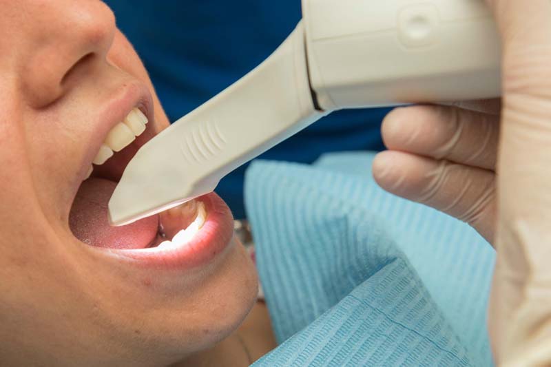
A 3D mouth scan changes that experience. Instead of asking you to bite into silicone or putty, the dentist moves a small handheld scanner around your teeth. Images appear on a screen as the device builds a detailed digital model of your mouth.
The result is not simply a more modern-looking appointment. Digital scanning can improve communication, make records easier to review and create a more direct route from assessment to personalised treatment planning.
What happens during a 3D mouth scan?
An intraoral scan is taken with a small camera-like device that captures the visible surfaces of the teeth and gums. As the scanner moves around the mouth, specialist software joins the images together to create a three-dimensional model.
You do not need to remain perfectly still while a tray of material hardens. If one small area has not been captured clearly, the dentist can return to it without repeating the entire process.
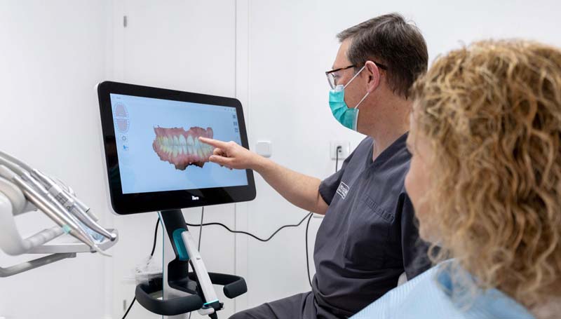
The model can then be enlarged, rotated and examined on screen. This gives the clinician a practical record of tooth shape, alignment and bite relationships while allowing the patient to see details that would be difficult to understand using a mirror alone.
It is also important to distinguish an intraoral scan from a CBCT scan. An intraoral scanner uses optical imaging to record the surfaces inside the mouth. A CBCT machine produces three-dimensional X-ray images of structures such as bone and roots. Depending on the treatment, a dentist may use one technology or combine both.
Precision is the key to comfort
Comfort in dentistry is not only about what happens in the chair. It is also about avoiding unnecessary repetition, poorly fitting appliances and appointments that could have been planned more clearly.
With conventional impressions, movement, moisture or distortion in the material can sometimes affect the model. A digital scan gives the clinician an immediate opportunity to inspect the record before the appointment ends. If something is missing, that area can usually be rescanned straight away.
Research comparing digital and conventional impressions has generally found that patients prefer digital scanning, particularly because it reduces the sensations associated with bulky trays and impression material. However, accuracy still depends on the scanner, the clinical situation and the experience of the operator.
Patients who want to understand the chairside experience in greater detail can read 3D intraoral scan: no molds, no discomfort, which explains how the digital model is captured and used at Condor Dent.
From a scan to digital planning dentistry
The scan is only the beginning. Its real value appears when the digital model becomes part of a wider treatment workflow.
In digital planning dentistry, the clinician can use the model to assess spacing, examine the bite and communicate with dental laboratories or aligner manufacturers. It may support the design of crowns, bridges, veneers, retainers and clear aligners, depending on the patient’s needs.
This does not mean software makes the clinical decisions. Technology organises and displays information, but the dentist must still examine the mouth, diagnose any problems and decide whether a proposed treatment is appropriate.
For a broader explanation of how scanning, computer-assisted design and three-dimensional imaging work together, explore digital dentistry in London and how 3D technology is changing smiles.
How digital scanning speeds up your aligner delivery
Traditional impressions must be checked, packaged and physically transported or converted into a digital format before an aligner plan can be created. A digital scan can be reviewed and transferred electronically, removing several manual stages from the workflow.
That does not guarantee that aligners will arrive immediately. The dentist must still complete the diagnosis, approve the proposed movements and allow time for manufacturing. Nevertheless, sending an accurate digital file can reduce avoidable delays caused by damaged impressions, unclear margins or models that need to be repeated.
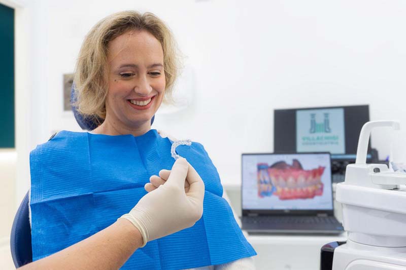
This is particularly relevant to Angel Aligner technology. The scan provides the digital starting model used to design a sequence of personalised aligners. Each stage represents a planned step in tooth movement, while review appointments allow the clinician to check whether the teeth are responding as expected.
Digital systems can make production more efficient, but the speed of delivery should never take priority over a complete orthodontic assessment.
Visualising your results before treatment starts
One of the most engaging parts of a digital appointment is seeing the proposed direction of treatment on screen.
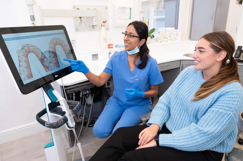
For orthodontic patients, software may simulate how tooth positions could change during the planned sequence. For restorative work, the digital model may help explain space, proportions or how a new restoration could relate to neighbouring teeth.
This preview is useful for communication, but it is not a photographic promise. Teeth respond biologically, and treatment plans sometimes require modifications. Gum condition, wear habits, compliance and individual healing can all influence the final result.
The real advantage is that patients can ask better questions. Instead of agreeing to an abstract description, they can discuss visible features of their own mouth and understand why particular steps have been recommended.
Why Condor Dent invests in high-tech machinery
Modern equipment is valuable when it improves the patient journey rather than simply adding impressive screens to the surgery. Condor Dent invests in digital infrastructure to support:
● Clearer visual explanations during consultations.
● Detailed records for treatment planning.
● More efficient communication with dental laboratories.
● Personalised production of aligners and restorations.
● Easier comparison of changes between appointments.
● Coordination between intraoral scans and other diagnostic images.
● Less dependence on messy physical impression materials.
Condor Dent also highlights access to in-house facilities, including X-ray equipment, a clinical operating environment and dental laboratory support, helping different stages of care remain connected.
Patients comparing the best dental clinics with the latest technology should look beyond the list of machines. The more useful questions are how the technology will be used, who interprets the information and whether it meaningfully improves diagnosis, communication or treatment delivery.
Does digital scanning mean painless dental care?
People often search for painless dental treatment, but no responsible dentist should promise that every procedure will be completely free from sensation.
The intraoral scan itself does not involve drilling, injections or impression material setting inside the mouth. Many patients therefore find it easier than conventional moulds. You may still feel the scanner tip moving around the teeth, particularly near the back of the mouth, and short pauses may be needed.
More importantly, a comfortable scan does not mean the treatment planned from it will be sensation-free. An implant, tooth preparation or orthodontic movement has its own clinical considerations. Digital technology may support precision and reduce some avoidable inconvenience, but pain management must always be discussed according to the actual procedure.
A better first step, not a replacement for the dentist
Perhaps the greatest change created by intraoral scanning is not technical. It changes the conversation.
When patients can see their own digital model, dental terminology becomes less remote. Crowding, wear, gaps and bite relationships are easier to point out. The appointment becomes a shared review rather than a one-sided explanation.
That does not remove the need for professional judgement. A scanner records surfaces; it does not independently diagnose decay, gum disease or every problem beneath the teeth. Clinical examination and any necessary X-rays remain essential.
Used properly, the 3D mouth scan is not a shortcut. It is a clearer starting point for well-planned care.
Frequently asked questions
Is a 3D mouth scan an X-ray?
No. An intraoral scanner uses optical technology to capture the visible surfaces of the teeth and gums. A CBCT scan is a separate type of three-dimensional X-ray used when information about roots, bone or deeper structures is required.
How long does an intraoral scan take?
The time varies according to the area being recorded and the complexity of the mouth. A partial scan may be relatively quick, while a full upper arch, lower arch and bite record will take longer.
Can I have a scan if I have a strong gag reflex?
Many people with a sensitive gag reflex find digital scanning easier than traditional impression trays. Tell the dental team in advance so they can work in shorter sections and allow pauses where necessary.
Can a 3D scan be used for Angel Aligner?
Yes, when the patient is clinically suitable for clear aligners. The digital model can support orthodontic planning and the manufacture of personalised trays, but a complete assessment is still necessary.
Are silicone dental moulds no longer used?
They are still useful in some situations. Digital scanning has replaced conventional impressions in many workflows, but the dentist may recommend a physical impression when it is more appropriate for a particular clinical case.

